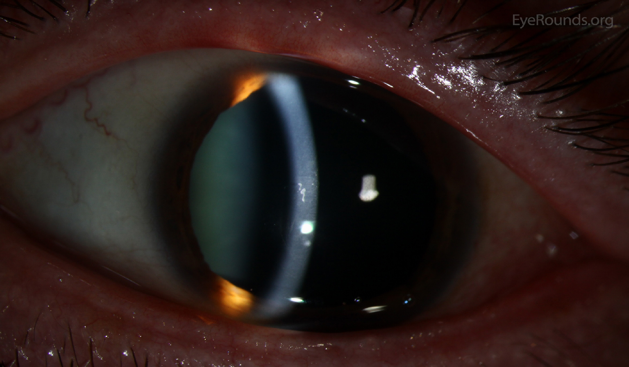
Epithelial Basement - Map Dot Fingerprint Dystrophy / Epithelial basement membrane dystrophy (ebmd) is the most common form of corneal dystrophy.. Epithelial basement membrane dystrophy (ebmd) is the most common type of corneal dystrophy, affecting 2% of the population.1 although the majority of patients remain asymptomatic or experience minor episodic subjective discomfort, about 10% will eventually complain of recurrent erosions and/or. Generally, this type of epithelium is found inside the body probably due to the fragile nature and forms the lining of the body cavities, blood and lymph vessels, heart and respiratory. Part b courtesy of christopher j. Images of epithelial basement membrane dystrophy. The main pathological feature of the disease is thickened, multilaminar and disfigured basement membrane of corneal epithelium.
The main pathological feature of the disease is thickened, multilaminar and disfigured basement membrane of corneal epithelium. Corneal anterior basement membrane dystrophy: Characteristic features of epithelial cells basement membranes intercellular adhesion & other junctions specializations of the apical cell surface microvilli stereocilia cilia types of basement membranes. Epithelial basement membrane dystrophy was initially undetected in three patients (cases 3, 4, and 7) in whom dystrophic changes were recognized postoperatively. These membranes serve as linings and covering for various body structures, and they also form glands.
Lamina Propria Wikipedia from upload.wikimedia.org Simple epithelium has only one cell layer where every cell is in direct contact with the underlying basement membrane. It is sometimes included in the group of corneal dystrophies. Epithelial basement membrane dystrophy is also sometimes referred to as either of the following: Characteristic features of epithelial cells basement membranes intercellular adhesion & other junctions specializations of the apical cell surface microvilli stereocilia cilia types of basement membranes. • type of epithelia • adhesion between epithelial cells • basement membrane • epithelial cell polarity • cell renewal. Images of epithelial basement membrane dystrophy. The spaces, surfaces and the cavities of the body. Along with other changes that occur among the epithelial cells, these changes give an appearance of maps, dots, and fingerprints within the cornea.
It is believed some 2% of the population is affected by it.
Epithelial basement membrane sits on. Epithelia are a collection of cells that form functional unit. The main pathological feature of the disease is thickened, multilaminar and disfigured basement membrane of corneal epithelium. Generally, this type of epithelium is found inside the body probably due to the fragile nature and forms the lining of the body cavities, blood and lymph vessels, heart and respiratory. Epithelial map changes can be obvious (a) or more subtle (b). It is sometimes included in the group of corneal dystrophies. • type of epithelia • adhesion between epithelial cells • basement membrane • epithelial cell polarity • cell renewal. Under the microscope, a structure called the epithelial basement membrane is abnormal. These membranes serve as linings and covering for various body structures, and they also form glands. Residents and fellows contest rules | international ophthalmologists contest rules. Since the normal corneal stroma is. The basement membrane consists of the basal lamina produced by epithelial cells, and a more fibrous reticular layer which is below the basal lamina. In 1964, it is also known as cogan's microcystic corneal dystrophy.
The basement membrane consists of the basal lamina produced by epithelial cells, and a more fibrous reticular layer which is below the basal lamina. Part b courtesy of christopher j. Corneal anterior basement membrane dystrophy: Generally, this type of epithelium is found inside the body probably due to the fragile nature and forms the lining of the body cavities, blood and lymph vessels, heart and respiratory. Epithelial map changes can be obvious (a) or more subtle (b).
Map Dot Fingerprint Dystrophy from webeye.ophth.uiowa.edu Epithelial basement membrane dystrophy is also sometimes referred to as either of the following: Epithelial cell in stool normal. Epithelial basement membrane dystrophy (ebmd), is a disorder of the eye that can cause pain and dryness. The main pathological feature of the disease is thickened, multilaminar and disfigured basement membrane of corneal epithelium. Residents and fellows contest rules | international ophthalmologists contest rules. Epithelial basement membrane sits on. It's transitional epithelium transitional epithelium is a stratified epithelium in which the shape of the surface cells changes (undergoes transitions) depending on the degree of stretch. Epithelial tissue and basement membranes.
Residents and fellows contest rules | international ophthalmologists contest rules.
Epithelial basement membrane sits on. Epithelial basement membrane dystrophy (ebmd), is a disorder of the eye that can cause pain and dryness. Since the normal corneal stroma is. Along with other changes that occur among the epithelial cells, these changes give an appearance of maps, dots, and fingerprints within the cornea. Residents and fellows contest rules | international ophthalmologists contest rules. In fact, for >20 years now, cultured human skin has been used as a source of new skin epithelial stem cells represent a ripe target for research into the fundamental mechanisms underlying these important processes. Epithelial membranes are comprised of epithelial tissue and connective tissue. Intercellular adhesion & other junctions. Since it was first described by cogan et.al. Epithelial cells line the surfaces and cavities of body tissues and organs. Epithelial basement membrane dystrophy is also sometimes referred to as either of the following: It is sometimes included in the group of corneal dystrophies. Stratified, but actually each cell is anchored to the basement the respiratory membrane consists of the epithelial cells of the alveolus, the endothelial cells of the capillary, and the two fused basement.
When a transitional epithelium is not stretched (for exampl. Along with other changes that occur among the epithelial cells, these changes give an appearance of maps, dots, and fingerprints within the cornea. It is sometimes included in the group of corneal dystrophies. Two primary types of epithelial membranes exist: All organs contain epithelia in some form.
Recurrent Corneal Erosions And Ptk from image.slidesharecdn.com Two primary types of epithelial membranes exist: Epithelial map changes can be obvious (a) or more subtle (b). Arrows show geographic map lines. The cells in epithelial tissue are tightly packed together with very little intercellular matrix. All organs contain epithelia in some form. Epithelia are a collection of cells that form functional unit. Epithelial basement membrane dystrophy is by far the most common epithelial dystrophy, but other dystrophies that can involve the corneal epithelium also cause pain or discomfort that can be relieved with soft lenses. 1) a thin extracellular supporting layer that separates a layer of epithelial cells from the underlying lamina propria and is composed of the basal lamina and reticular lamina.
Epithelial map changes can be obvious (a) or more subtle (b).
Characteristic features of epithelial cells basement membranes intercellular adhesion & other junctions specializations of the apical cell surface microvilli stereocilia cilia types of basement membranes. In 1964, it is also known as cogan's microcystic corneal dystrophy. In the remaining three patients (cases 1, 8, and 9), the dystrophy was judged to be insignificant in its appearance and by its paucity of symptoms. Epithelial basement membrane dystrophy is also sometimes referred to as either of the following: Epithelial cells line the surfaces and cavities of body tissues and organs. Epithelial basement membrane dystrophy is by far the most common epithelial dystrophy, but other dystrophies that can involve the corneal epithelium also cause pain or discomfort that can be relieved with soft lenses. In fact, for >20 years now, cultured human skin has been used as a source of new skin epithelial stem cells represent a ripe target for research into the fundamental mechanisms underlying these important processes. Is degenerated leiomyoma associated with increased epithelial antigen? 1) a thin extracellular supporting layer that separates a layer of epithelial cells from the underlying lamina propria and is composed of the basal lamina and reticular lamina. Two primary types of epithelial membranes exist: Epithelia are a collection of cells that form functional unit. The cells in epithelial tissue are tightly packed together with very little intercellular matrix. These membranes serve as linings and covering for various body structures, and they also form glands.